
ML387 : HPGD (15-hydroxyprostaglandin dehydrogenase [NAD(+)]) Inhibitor
ML387
Target Name
15-hydroxyprostaglandin dehydrogenase [NAD(+)]
Target Alias
HPGD
Target Class
Dehydrogenase
Mechanism of Action
Inhibitor of HPGD
Biological / Disease Relevance
Inflammation, Differentiation, Cellular signaling, Human NAD+ dependent 15-hydroxyprostaglandin dehydrogenase
In vitro activity
HPGD bioassay (IC50)Cellular activity
PGE2 Assay in A549 (IC50)Cellular activity
PGE2 Assay in LNCaP (IC50)Target Information
The central role of hydroxyprostaglandin dehydrogenase (HPGD) is to inactivate prostaglandins such as prostaglandin E (PGE ) or prostaglandin D (PGD ). These prostaglandins are involved in a number of processes, including inflammation, differentiation, and cellular signaling, which makes HPGD an important target for pharmacological intervention. High throughput screening of the Molecular Libraries Small Molecule Repository (MLSMR) led to the identification of three small molecule inhibitors of HPGD with differing mechanisms of action (ML147, ML148, and ML149). Herein, we report, the structure-activity relationship (SAR) studies, cell-based characterization and selectivity studies around the ML148 and ML149 scaffolds. These combined efforts led to the identification of two novel small molecule chemical probes (ML387 and ML388) with nanomolar inhibitory activity and excellent selectivity toward HPGD. Moreover, these compounds were shown to potently promote PGE production in both A549 lung cancer and LNCaP prostate cancer cells. As part of these studies, we also profiled top compounds for select in vitro ADME properties to facilitate future optimization efforts.
Properties
ML387
NCGC00262228
| Physical & chemical properties | ||||
|---|---|---|---|---|
| Molecular Weight | 335.4 g/mol | |||
| Molecular Formula | C20H21N3O2 | |||
| cLogP | 3.4 | |||
| PSA | 47.4 Ų | |||
| Storage | ||||
| Solubility | ||||
| CAS Number | ||||
SMILES:
COC1=CC=C(N2C=NC3=CC(C(N4CCCCC4)=O)=CC=C23)C=C1
InChI:
1S/C20H21N3O2/c1-25-17-8-6-16(7-9-17)23-14-21-18-13-15(5-10-19(18)23)20(24)22-11-3-2-4-12-22/h5-10,13-14H,2-4,11-12H2,1H3
InChIKey:
FUCWZIFGSAFUAO-UHFFFAOYSA-N
Activity
Summary activity statement /
Prostaglandins play central roles as signaling molecules, hence their levels are tightly regulated. One approach to study prostaglandins deregulation is through the modulation of their metabolism. HPGD is central to the metabolism of three key prostaglandins: PGE , PGD , and 15d-PGJ . The two reported probes, ML387 (SID 162211043; CID 3474778) and ML388, are small molecules inhibitors of HPGD with nanomolar potency, excellent selectivity versus five other dehydrogenases, and improved ADME properties when compared to the original probes. Moreover, their efficacy was demonstrated at the cellular level through induction of PGE production in A549 (lung cancer) cells and LNCaP prostate cancer cells. Given these attributes, ML387 and ML388 would be useful for studying the effect of pharmacological inhibition of HPGD in diseases such as cancer (e.g. cell migration, proliferation) and wound healing (prevention of excessive scar tissue).
In vitro activity - Selectivity Assay
| Bioassay | ML387 (IC50) |
|---|---|
|
HPGD |
0.019 uM |
|
ALDH1A1 (Anti-Target) |
1358 uM |
|
LDHA (Anti-Target) |
Inactive |
|
LDHB (Anti-target) |
Inactive |
|
rGAPDH |
Inactive |
|
HSD17B4 |
Inactive |
Summary /
The inhibitory activities of ML387 versus a panel of dehydrogenases is assessed. The probe is found to have 71 fold selective against HPGD vs. ALDH1A1 and > 1000 fold vs. other dehydrogenases.
In vitro activity - ADME profiling
| ML387 | |
|---|---|
|
PAMPA Permeability (pH 7.4) |
624 x 10^-6 cm/s |
|
Aqueous Kinetic Solubility in PBS (pH 7.4) |
> 50 uM |
|
Rat Liver Microsomal Stability with NADPH (T1/2) |
23 min |
Summary /
PAMPA: Parallel Artificial Membrane Permeability Assay; the assay measures the passive transport of the probes through a lipid bilayer. The probes showed no degradation without NADPH over a 1 h period.
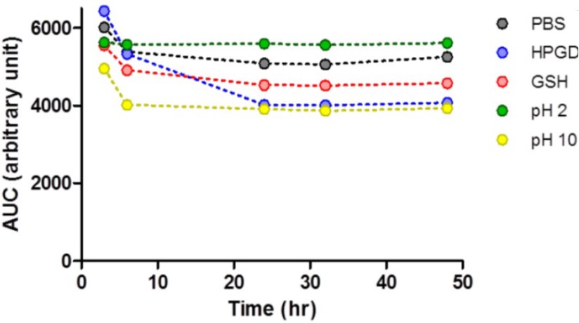
Figure 1. Stability of ML387 over time in aqueous solution at room temperature, as measured by the area under the curve (AUC) of the probe HPLC signal observed at 254 nm. PBS: phosphate buffer saline, pH 7.4; HPGD: assay buffer; GSH: 10 mM GSH in PBS buffer.
References
- Extended Characterization of HPGD Inhibitors: Activity in Primary Assay (HPGD)
- Duveau DY, Yasgar A, Hu X, et al. Discovery of two small molecule inhibitors, ML387 and ML388, of human NAD+-dependent 15-hydroxyprostaglandin dehydrogenase. 2013 Dec 15 [Updated 2015 Feb 11]. In: Probe Reports from the NIH Molecular Libraries Program [Internet]. Bethesda (MD): National Center for Biotechnology Information (US); 2010-. Available from: https://www.ncbi.nlm.nih.gov/books/NBK280041/