
ML290 : RXFP1 (Relaxin Receptor 1) Agonist

ML290
Target Name
Relaxin Receptor 1
Target Alias
RXFP1
Target Class
G-protein Coupled Receptor
Mechanism of Action
Agonist of RXFP1
Biological / Disease Relevance
Acute Heart Failure (AHF), Recombinant Human Relaxin Therapy Alternative, RXFP1 activation pathway
In vitro activity
RXFP1 bioassay (AC50)Cellular activity
VEGF assay (IC50)Target Information
The peptide hormone relaxin has been clinically investigated as a beneficial treatment for acute heart failure (AHF). Relaxin has been shown to reduce blood pressure and promote vascular compliance in clinical studies, in addition to being able to remodel heart lesions. The target of relaxin’s action is the class B G-protein coupled receptor RXFP1. Here we present the discovery of the first small molecule agonists of RXFP1 disclosed in the literature. Optimized compounds from this series are potent and highly selective RXFP1 agonists with similar efficacy as the natural hormone in functional assays. These molecules are easy to synthesize and the represented analog ML290 showed excellent in vitro absorption, distribution, metabolism, and excretion (ADME) data and in vivo pharmacokinetic (PK) properties. From our studies, we conclude that this probe, ML290, should be a very useful tool for the study of RXFP1 activation in pre-clinical disease models of heart failure and other diseases, and might provide a lead for the development of a small-molecule drug as an alternative to the current expensive recombinant human relaxin therapy.
Properties
ML290
NCGC00250135
| Physical & chemical properties | ||||
|---|---|---|---|---|
| Molecular Weight | 506.5 g/mol | |||
| Molecular Formula | C24H21F3N2O5S | |||
| cLogP | 5.4 | |||
| PSA | 110 Ų | |||
| Storage | ||||
| Solubility | ||||
| CAS Number | 1482500-76-4 | |||
SMILES:
CC(OC1=C(C(NC2=C(C(NC3=CC=CC(S(=O)(C(F)(F)F)=O)=C3)=O)C=CC=C2)=O)C=CC=C1)C
InChI:
1S/C24H21F3N2O5S/c1-15(2)34-21-13-6-4-11-19(21)23(31)29-20-12-5-3-10-18(20)22(30)28-16-8-7-9-17(14-16)35(32,33)24(25,26)27/h3-15H,1-2H3,(H,28,30)(H,29,31)
InChIKey:
RSYHJSDOGMSLDH-UHFFFAOYSA-N
Activity
Summary activity statement /
The probe molecule, ML290 (SID 134225125; CID 56593349), presented in this report is a potent and highly selective RXFP1 agonist. The molecule can be used in cell-based studies to investigate the role of RXFP1 activation in downstream signaling events, including VEGF elevation, MAP kinase activation, NO production, and decreases in the production of inflammatory cytokines, such as TNF-α and TGF-β. In addition, ML290 is metabolically stable, with very good microsomal and plasma stability, reasonable physical properties and excellent in vivo pharmacokinetics, making it a good candidate for further in vivo animal disease model efficacy studies.
In vitro activity - Selectivity and Cytotoxicity Assay
| Bioassay | ML290 (IC50) |
|---|---|
|
RXFP1 |
94 nM |
|
RXFP2 (Anti-Target) |
< 30%, 1.5 uM |
|
V1b (Anti-target) |
Inactive |
Summary /
ML290 is found to have > 15 fold selective against RXFP1 vs. RXFP2.
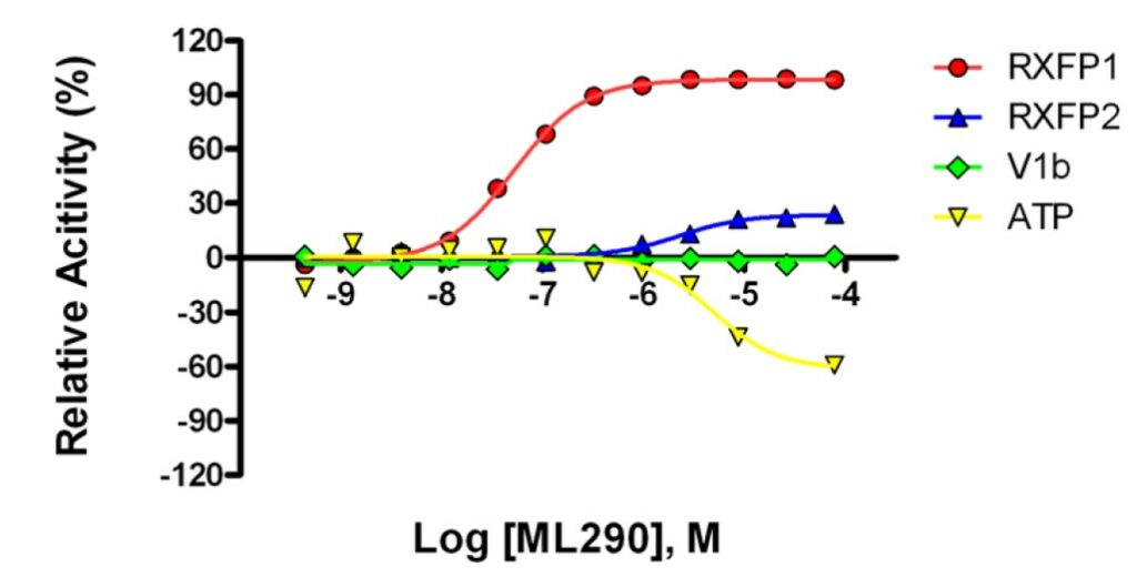
Figure 1. Graphical representation of the dose response curves of ML290 in RXFP1 (red), RXFP2 (blue), V1b cell (green) lines, and in ATP cytotoxicity assay (yellow). RXFP1, RXFP2, and V1b data were all read at 30 minutes, while ATP data was read at 72 hours.
Cellular activity - Functional Assay: VEGF stimulation in THP1-RXFP1 cells
Summary /
THP1 cells (human acute monocytic leukemia cell line) were used to analyze the stimulation of VEGF gene expression after treatment with relaxin or compounds. The VEGF stimulation in these cultured endometrial cells is a well-established property of relaxin (Unemori 1999, Unemori 2000). This effect is most probably responsible for the observed angiogenic and neovascularization properties of relaxin in various settings.20 400,000 THP1 cells (0.4 mL at 1×10⁶ cells/mL) in test media (RPMI-1640 without phenol red, 0.5% FBS, 1x Pen/Strep, 0.05 mM of 2-mercaptoethanol) were seeded in each well on a 24-well plate. After 24 hours incubation at 37 °C, 5% CO2, relaxin or compounds were added for 2 hours. The cells were harvested and RNA was extracted by the Trizol (Invitrogen, Carlsbad, CA) method according to manufacturers’ instructions. cDNA was synthesized by using Verso cDNA kit (Thermo Scientific, Waltham, MA) according to manufacturer’s protocol. Quantitative real time RT-PCR for VEGF and GAPDH gene expression was done using a Roche LightCycler 480 (Roche Diagnostics, Indianapolis, IN) with the appropriate set of primers and probes spanning different exons. The relative fold change in VEGF mRNA level was calculated by the comparative Ct (2–ΔΔCt) method using GAPDH expression for normalization of RNA.
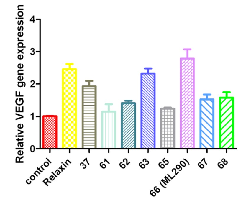
Figure 2. Activation of VEGF expression in THP1-RXFP1 cells by relaxin at 10 ng/mL and eight selected analogs at 250 nM. All analogs but 61 show significant (p<0.01) up-regulation of VEGF expression.
Cellular activity - Functional assay: Cell Impedance
Summary /
It was previously shown that relaxin increases cell impedance in RXFP1 transfected cells (Shemesh 2009). Cell-substrate impedance was measured using a Roche DP RCTA xCELLigence Analyzer (Roche Diagnostics, Indianapolis, IN) on E-Plates. Real Time Cell Analyzer (RTCA) allows for continuous time-resolved measurement of cellular index without additional labeling. Cell number, cellular adherence to the plate, and intracellular interactions all contribute to the total cellular impedance. The effect of the compound treatment is only measured within the first hour, changes in cellular density are unlikely to contribute to the overall effect, and therefore cellular impedance is most likely caused by intercellular interactions, or signaling. Cell Index (CI) was calculated by subtracting impedance at the beginning of experiment Z0 from impedance at each individual time point Zt, divided by 15Ω [CΙt=( Z0-Zt)/15 Ω]. Delta Cellular Indices were calculated as the change of impedance at a given time t, from the time of compound addition (CIcompound) ΔCIt=CIt-CIcompound. Impedance at each time point was then normalized to the average of quadruplicate CI of cells treated with vehicle (V1, V2, V3, and V4), to calculate normalized delta Cell Index NΔCI= (CIt-CIcompound) /Average[ΔCIV1, ΔCIV2, ΔCIV3, ΔCIV4]. Maximal relaxin activity was assigned a value of 100% and all other values adjusted proportionally. The cell line stably transfected with RXFP1 receptor HEK293-RXFP1 was used for cell impedance assay to confirm relaxin-like properties of the compounds. To equilibrate the plates, 100 μL of test media (DMEM, 1% FBS, 1× Pen/Strep) was added to each well of E-Plate (Roche Diagnostics, Indianapolis, IN) and the plate was incubated at room temperature for 30 minutes at which point baseline impedance was measures. Then 20,000 HEK293-RXFP1 cell or HEK293 cells (parental control cell line) were added per well in a volume of 100 μL test media and allowed to sediment at room temperature for 30 minutes. The plate was placed into xCELLigence RTCA DP Instrument in the CO2 incubator overnight to allow the cells to attach. Relaxin (10 ng/mL), vehicle, or compounds at different concentrations (250, 500, and 750 nM) were added to the wells and the cellular impedance was measured every 10-30 seconds for 1 hour.
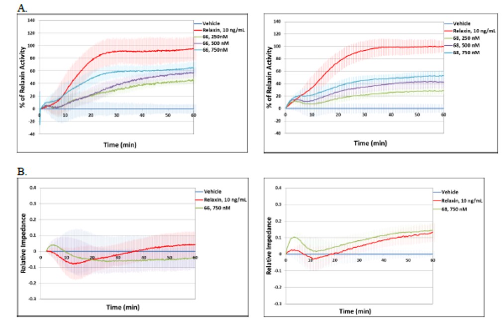
Figure 3. A) Effect of relaxin at 10 ng/mL, compared to 66 (ML290) and 68 at 250 nM, 500 nM, and 750 nM on cell impedance in HEK293-RXFP1 cells. The data were normalized to maximal cell index of relaxin treated cells (100%). B) Effect of relaxin at 10 ng/mL, compared to 66 (ML290) and 68 at 750 nM on cell impedance in parental HEK293 cells.
In vitro and vivo - ADME and PK Profiling
Summary /
ML290 (compound 66) showed a promising ADME profile and excellent in vivo pharmacokinetics property.
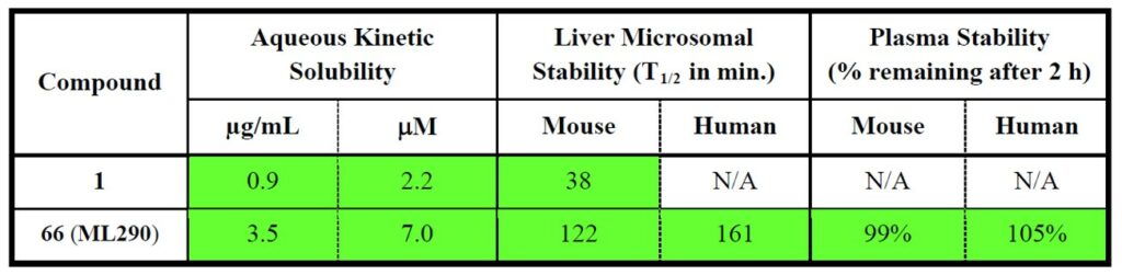
Table 1. Comparison of ADME profile for hit 1 and ML290.
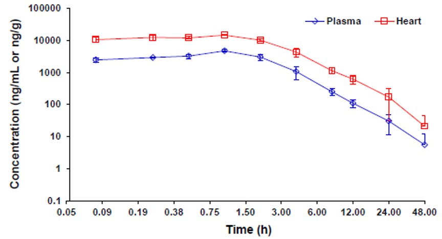
Figure 4. Mean plasma and heart concentration-time profiles of ML290 after an IP dose of 30 mg/kg in male C57BL/6 mice (N = 3). No abnormal clinical observation was found during the in life phase. The IP dosing solution was prepared in 10% NMP + 10% Solutol HS15 + 10% PEG400 + 70% Saline. Full PK parameters are listed in the Appendix Table A1 and A2.
References
- qHTS Assay for Agonists of the Relaxin Receptor RXFP1: Summary
- Xiao J, Chen CZ, Huang Z, et al. Discovery, optimization, and biological activity of the first potent and selective small-molecule agonist series of human relaxin receptor 1 (RXFP1) 2012 Mar 10 [Updated 2013 May 8]. In: Probe Reports from the NIH Molecular Libraries Program [Internet]. Bethesda (MD): National Center for Biotechnology Information (US); 2010-. Available from: https://www.ncbi.nlm.nih.gov/books/NBK153218/
- Unemori EN, Erikson ME, Rocco SE, et al. Relaxin stimulates expression of vascular endothelial growth factor in normal human endometrial cells in vitro and is associated with menometrorrhagia in women. Hum Reprod. 1999;14(3):800-806. doi:10.1093/humrep/14.3.800
- Unemori EN, Lewis M, Constant J, et al. Relaxin induces vascular endothelial growth factor expression and angiogenesis selectively at wound sites. Wound Repair Regen. 2000;8(5):361-370. doi:10.1111/j.1524-475x.2000.00361.x
- Shemesh R, Hermesh C, Toporik A, et al. Activation of relaxin-related receptors by short, linear peptides derived from a collagen-containing precursor. Ann N Y Acad Sci. 2009;1160:78-86. doi:10.1111/j.1749-6632.2009.03827.x